What are major Factors in Stroke Recovery and Future Directions of Therapy.
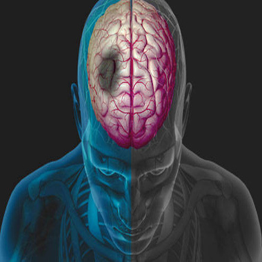
Stroke is the fourth leading cause of death and a leading cause of long-term disability in the United States. The economic burden of stroke is huge. As of 1990, the average lifetime cost of caring for one stroke patient was estimated at $103,576, including care costs for all phases (acute treatment, rehabilitation, ambulatory, and nursing home).
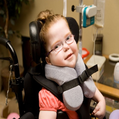
With improvement in acute stroke care and the establishment of stroke centers nationwide, more stroke victims survive, but with varying degrees of disability. The common deficits associated with stroke are motor impairment (including limb spasticity), sensory impairment, language impairment (aphasia and/or dysarthria), dysphagia, cognitive impairment, visual impairment, and post-stroke depression. Motor impairment is the most common physical deficit. Although the demand for effective and accessible rehabilitation therapies has tremendously increased over time, only a limited set of effective neuro-rehabilitative modalities are currently available while a few promising therapeutic options are still in research stages.
Mechanisms of Recovery
The first recovery mechanism is resolution of harmful local factors, which generally accounts for early spontaneous improvement after stroke (usually within the first 3-6 mo). These processes include resolution of local edema, resorption of local toxins, improvement of local circulation, and recovery of partially damaged ischemic neurons.
The second recovery mechanism, which may continue for many months, is neuroplasticity, which can take place early or late. Brain plasticity is the ability of the nervous system to modify its structural and functional organization. The 2 most plausible forms of plasticity are collateral sprouting of new synaptic connections and unmasking of previously latent functional pathways.
Other mechanisms of plasticity include assumption of function by undamaged, redundant neural pathways, reversibility from diaschisis, denervation supersensitivity, and regenerative proximal sprouting of transected neuronal axons. Experimental evidence indicates that plasticity can be altered by several external factors, including pharmacologic agents, electrical stimulation, and environmental stimulation.
A key aspect of neuro-plasticity that has important implications for rehabilitation is the fact that the modifications in neuronal networks are use-dependent. Animal experimental studies and clinical trials in humans have shown that forced use and functional training contribute to improved function. On the other hand, techniques that promote non-use may inhibit recovery.
Major Determinants in the Stroke Recovery Process
Stroke affects everybody differently. Many stroke survivors continue to improve over a long time, sometimes over a number of years. Recovery from stroke involves making changes in the physical, social and, emotional aspects of your life. You will make changes to prevent additional strokes as well as to facilitate your life-long recovery.
It is normal to feel angry, anxious or depressed after a stroke. You may feel worried about work, money and relationships, and the tiredness caused by stroke can make things worse.
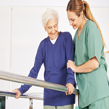
Rehabilitation is about getting back to normal life and living as independent a life as possible. It involves taking an active approach to ensure that your life goes on. This can mean learning new skills or relearning old ones. It may involve adapting to new limitations and post-stroke conditions. Or it can mean finding new social, emotional, and practical support to live your best life post-stroke.
Stroke recovery is a sophisticated biological process with many factors influencing the recovery trajectory. Rehabilitative strategies aiming to improve post-stroke recovery outcomes require a comprehensive understanding of those factors. The recent advances in imaging tools, neuro-physiological methods, and genetics have increased our knowledge about the post-stroke recovery process. In this section, we will summarize the major determinants of recovery process, including:
Initial Injury
This is the most important prognostic factor for subsequent stroke recovery. Neuroimaging findings to know the extent of initial injury including stroke size and location are an important adjunct to the neurologic exam when gauging prognosis. Early after stroke, the neurologic exam alone can suggest a falsely grim or favorable prognosis. For example, a patient may have a small stroke on neuroimaging and present with stupor or coma caused by seizure or metabolic derangement that is reversible. Conversely, a patient presenting with mild stroke and a low NIHSS (National Institutes of Health Stroke Scale) score on examination may have large vessel occlusion and a large perfusion deficit on neuroimaging, suggesting the possibility of stroke progression and worse outcome.
Infarct Volume
The volume of acute infarction on neuroimaging studies may be used to estimate stroke outcome. In one small study, the volume of ischemic tissue determined by diffusion-weighted MRI within 36 hours of stroke onset combined with the NIHSS score and time from stroke onset to imaging predicted the functional outcome at three months better than any of the individual factors alone. A much larger study analyzed data from over 1800 patients who had CT or MRI within 72 hours of ischemic stroke onset and found that initial infarct volume was an independent predictor of stroke outcome at 90 days, along with age and NIHSS score. In these and most other reports, the vast majority of infarcts analyzed were supratentorial (eg, anterior circulation, middle cerebral artery territory) and the results may not apply to posterior circulation or infratentorial infarcts, in which an infarct of small volume can result in severe disability.
Infarct Location
The prognosis for stroke recovery may vary by the affected vascular territory and site of ischemic brain injury. For example acute occlusion of the cervical internal carotid artery, basilar artery, or a large intracranial artery is associated with an increased risk of poor outcome while strokes in the insular region (supplied by the insular branch of the middle cerebral artery) have been associated with increased mortality. Anterior choroidal artery infarctions may be more likely to progress in the first few days after stroke than other subtypes.
In addition to stroke volume and location, there are other features identifiable on neuroimaging that may suggest poor prognosis:
●Diffusion-perfusion mismatch
●Poor collateral blood flow
●Development of cerebral edema in nonlacunar ischemic stroke
●Acute corticospinal tract Wallerian degeneration
The etiology or mechanism of ischemic stroke could also influence prognosis for recovery.
●Patients with lacunar infarcts have a better prognosis up to one year after onset than those with infarcts due to other stroke mechanisms.
●Cryptogenic stroke, where no mechanism of stroke is identified, tends to have a better prognosis up to one year following onset.
●Patients with strokes of cardioembolic or large artery etiology tend to have worse prognosis for recovery compared with other ischemic stroke subtypes.
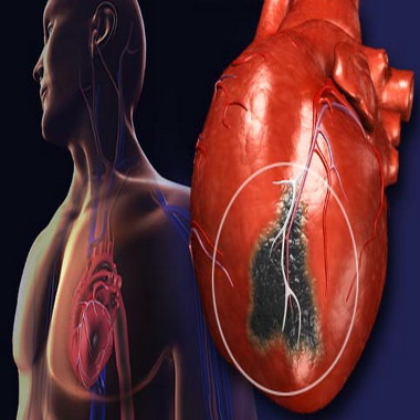
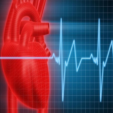
A host of pre-stroke co-morbid conditions are associated with an increased risk of poor outcome following ischemic stroke, including the following:
●Atrial fibrillation
●Cancer
●Coronary artery disease
●Dementia
●Dependency
●Diabetes mellitus
●Hyperglycemia (eg, blood glucose >6.1 mmol/L [>110 mg/dL]) on admission
●Heart failure
●Myocardial infarction
●Renal dysfunction or dialysis
●Poor nutritional status
●Low hemoglobin level
Post-Stroke Depression
Mood depression is a common and serious complication after stroke. According to epidemiological studies, nearly 30% of stroke patients develop depression, either in the early or in the late stages after stroke. Although depression may affect functional recovery and quality of life after stroke, such condition is often ignored.
Rehabilitation Therapeutics
The type, dosage, and timing of rehabilitation therapies play important roles in stroke recovery, but optimization of these parameters needs further refinement. For example, patients who suffered a stroke in the previous 3 to 9 months with some degree of wrist and finger movements can have a greater benefit from constraint-induced movement therapy (CIMT) over traditional therapy, but such therapy is only equally effective and not superior to customary therapy if it is given in the very acute phase.
Socio-demographic Determinants
Socio-demographic factors are important determinants of stroke recovery. Although some studies have shown that increased age is a significant prognostic factor for poorer outcomes, this remains controversial. Overall, the influence of age on recovery is probably minimal after adjusting for other factors.
Women are less likely to achieve functional independence and more likely to be disabled than men, but the mechanism for this difference is not clear. Racial disparity is also well documented in stroke. Blacks have a significantly higher stroke incidence, greater initial stroke severity, higher stroke mortality, and poorer recovery outcome compared with Whites.
Socioeconomic status (insurance coverage, educational level, household income, etc.) is tied with access issues and subsequent recovery outcomes. The relative or complete lack of health insurance coverage may delay or limit access to rehabilitative services and is likely associated with less optimal recovery.
Genetic Factors
Recovery of motor function after stroke involves relearning motor skills and is mediated by neuroplasticity. Recent research has focused on developing rehabilitation strategies that facilitate such neuroplasticity to maximize functional outcome poststroke. Although many molecular signaling pathways are involved, brain-derived neurotrophic factor (BDNF) has emerged as a key facilitator of neuroplasticity involved in motor learning and rehabilitation after stroke.
Recent advancements in the understanding of the role of brain-derived neurotrophic factor (BDNF) in neuroplasticity may provide important information for the development of new poststroke rehabilitation strategies. Brain-derived neurotrophic factor is a member of the neurotrophin family, a group of proteins involved in neuroprotection, neurogenesis, and neuroplasticity, and has been identified as a key mediator of motor learning and rehabilitation after stroke.
New areas of research are beginning to inform the development of rehabilitation strategies that take into account the importance of BDNF for motor recovery after stroke. These areas of research include consideration of aerobic exercise effects on brain function and the incorporation of genetic information to individualize therapy.
Rehabilitation strategies that optimize BDNF effects on neuroplasticity may be especially effective for improving motor function post-stroke. Two potential post-stroke rehabilitation strategies that consider the importance of BDNF are the use of aerobic exercise to enhance brain function and the incorporation of genetic information to individualize therapy. Converging evidence demonstrates that aerobic exercise increases BDNF production and consequently enhances learning and memory processes.
This perspective article discusses evidence that aerobic exercise promotes neuroplasticity by increasing BDNF production and considers how aerobic exercise may facilitate the acquisition and retention of motor skills for poststroke rehabilitation.
In a recent study on the impact of the BDNF gene val66met polymorphism on motor learning and response to rehabilitation it is concluded that the effects of aerobic exercise on BDNF and motor learning may be better exploited if aerobic exercise is paired more closely in time with motor training. Additionally, information about BDNF genotype could provide insight into the type and magnitude of effects that aerobic exercise may have across individuals and potentially help guide an individualized prescription of aerobic exercise to enhance motor rehabilitation post-stroke.
Future Directions of Stroke Recovery
New technologies are only just beginning to scratch the surface of what can be achieved in rehabilitation. Robotics, mechanized ambulation, virtual reality, and mental practice all enable the patient to meet the criteria of task-specific therapy.
These include robotics, constraint-induced movement therapy (CIMT), and pharmacological therapy such as levodopa, and selective serotonin reuptake inhibitors. In addition, several forms of non-invasive cortical stimulation such as rapid transcranial magnetic stimulation (rTMS), transcranial direct current stimulation (tDCS), and theta-burst stimulation (TBS) have shown promise in early phase studies.
One hurdle to implementation of restorative therapies is the heterogeneity of stroke, as injury location and size vary widely from one patient to the next. The ability to assign the right patients to the right therapies would maximize treatment effects, for example, by confirming the presence of a therapy’s biological target. Because cellular and molecular measurements are generally inaccessible in human subjects, a number of neuroimaging methods have been examined to better understand, predict, and guide post-stroke restorative therapies.
Let’s have a good look at some of these new strategies in stroke recovery.
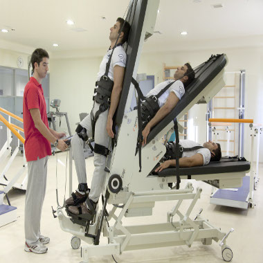
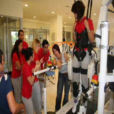
Robotic-Assisted Rehabilitation
In an attempt to supplement the use of therapists in providing a more intensive therapeutic environment, there has been increasing interest in the use of robotics in stroke rehabilitation.
Research into rehabilitation robotics has grown rapidly and the number of therapeutic rehabilitation robots has expanded dramatically during the last two decades. Robotic rehabilitation therapy can deliver high-dosage and high-intensity training, making it useful for patients with motor disorders caused by stroke or spinal cord disease. Robotic devices used for motor rehabilitation include end-effector and exoskeleton types which are useful for both the upper limbs and the lower limbs.
Both end-effector and the exoskeleton devices have proven to be effective complements to conventional physiotherapy in patients with subacute stroke, but there is no clear evidence that robotic gait training is superior to conventional physiotherapy in patients with chronic stroke or when delivered alone.
This advancement in technology has made robotics available for rehabilitation intervention. The most important advantage of using robot technology in rehabilitation intervention is the ability to deliver high-dosage and high-intensity training. This property makes robotic therapy a promising novel technology for rehabilitation of patients with motor disorders caused by stroke.
Virtual Reality and Motor Imagery
One of the newest potential therapies currently under study is virtual reality as a training tool in stroke rehabilitation. Virtual reality is computer technology that simulates real-life learning and allows for increased intensity of training while providing augmented sensory feedback. Hundreds of clinicians and caregivers around the world treat patients with gesture based virtual reality and interactive computer-based therapy systems that let patients have fun while stretching their physical and cognitive capabilities.
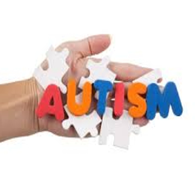
Immersive video technology projects a patient’s real-time image into exciting virtual environments. Patients perform clinician-prescribed exercises, games and activities, using body movement to control the program, so there’s no need to wear, hold or be attached to anything. Virtual reality exercise regimes are proven by research to deliver remarkable therapeutic benefits to physiotherapy patients. They also help those suffering from cerebral palsy, stroke, traumatic brain injury, autism and other conditions.
Patients experience improvement in trunk control, limb use, balance, mobility and gross and fine motor movement. They can also see enhanced cognitive and executive functioning and a greater sense of confidence and empowerment. Many patients find they require less surgical intervention and medication as a result of virtual therapy.
An even more interesting concept that is being used in rehabilitation is the concept of motor imagery, which is being incorporated into rehabilitation trials, particularly for motor retraining of hemiparetic limbs. It has been shown that motor imagery can result in increased functional Magnetic Resonance Image activity, a potential precursor of recovery. Stevens et al in 2 chronic hemiparetic stroke survivors used imagination and mirrors to trick patients’ brains into thinking that they were moving their arms and legs.
Patients underwent intensive training utilizing motor imagery consisting of imagined wrist movements and mental stimulations of reaching and object manipulation using a mirror box whereby they move their unaffected limbs in a mirror but which appears like they are moving their affected limb. In addition, patients were told to imagine that they were moving their paralyzed limb. Patients who used this technique showed improvement in the motor function of the paretic limb.
Botulinum toxin
Botulinum toxin is a neurotoxin produced by Clostridium botulinum, a bacterium that causes food poisoning (botulism). There are seven known types of C. botulinum toxin, but only types A; (BOTOX Cosmetic) and B (Myobloc) are used as medical treatments.
Botulinum toxin has proven to be useful in the treatment of many forms of dystonia, including the following:
• blepharospasm–forceful involuntary closure of the eyelids
• strabismus–misalignment of the eyes
• hemifacial spasm–sudden contraction of the muscles on one side of the face
• spasmodic torticollis, or cervical dystonia–muscle spasm in the neck that causes the head to turn to one side, and sometimes forward or backward
• oromandibular dystonia–continuous spasms of the face, jaw, neck, tongue, larynx, and in severe cases, the respiratory system
• urinary retention–severe inability to urinate that requires catheterization
• spasmodic dysphonia–spasm of the vocal cords that causes sudden disruption of speech
• stuttering–repetitions of parts of words and whole words, long pauses, elongated sounds
• voice tremor–quavering vocalization
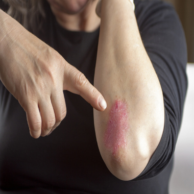
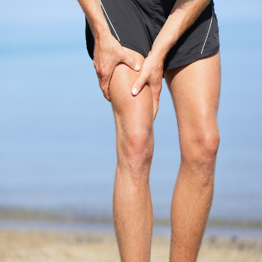
• limb spasticity (e.g., following stroke)
Constraint Induced Movement Therapy (CIMT)
The focus of CIMT is to combine restraint of the unaffected limb and intensive use of the affected limb. Types of restraints include a sling or triangular bandage, a splint, a sling combined with a resting hand splint, a half glove, and a mitt. Determination of the type of restraint used for therapy depends on the required level of safety vs. intensity of therapy.
Some restraints restrict the wearer from using their hand and wrist; though allow use of their non-involved upper extremity for protection by extension of their arm in case of loss of balance or falls. However, restraints that allow some use of the non-involved extremity will result in less intensive practice because the non-involved arm can still be used in complete tasks. This consists of an intensive upper limb exercise program, coupled with placing the unaffected arm in a restrictive mitt to encourage use of the weak upper limb. Classic CIMT includes six hours of supervised therapy five days per week for a two week period. Modified versions of CIMT may involve less intensive therapy programs over of a longer period of time.
Electrical stimulation
Electrical Stimulation of the nerves or muscles in the arm or leg can cause movement of the weak limb, and there is some evidence that repeated use of this treatment can result in some degree of restoration of movement. Functional Electrical stimulation devices used for the upper limb include the NESS H200 and the NeuromoveT™. Functional Electrical stimulation devices for the leg include the MyGait, NESS L300, and the WalkAide®. The lower limb devices are often used as a partial substitute for a plastic leg brace.
Partial body weight-supported treadmill training
This is a technique that uses a harness to reduce the amount of weight the stroke survivor is bearing through the legs, and combines this with practice walking on a treadmill. This technique appears most useful for stroke survivors who are early in their recovery and having more substantial difficulty in regaining walking ability. Treadmill therapy with partial body weight support system is a promising new approach to improve gait ability after stroke.
This task-specific approach enables non-ambulatory patients the repetitive practice of complex gait cycles instead of single-limb gait-preparatory maneuvers. Patients walk more symmetrically with less spasticity and better cardiovascular efficiency on the treadmill than with floor walking. Several controlled, clinical studies have shown the potential of treadmill training as a therapeutic intervention for non-ambulatory patients with chronic stroke-related hemiplegia. Furthermore, controlled trials in acute stroke survivors have shown that treadmill training is as effective as other physiotherapy approaches that stress the repetitive practice of gait.
Robotic therapy such as the Lokomat, Ambulator and Lite Gait® Partial-Weight-Bearing Gait Therapy Device represents an attempt to automate this type of therapy.
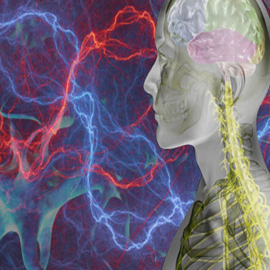
Brain Stimulation
Brain stimulation therapies involve activating or touching the brain directly with electricity, magnets, or implants to treat depression and other disorders. Brain stimulation using trans-cranial magnetic stimulation, also known as TMS and trans-cranial direct current stimulation (TDCS) are both experimental therapies that may enhance the brains ability to rewire itself after a stroke.
With the availability of these myriads of resources and equipment, neuro-rehabilitation and stroke recovery will help a lot of patients to recover functional use of their limbs and resume activities of daily living and the lifestyles they are used to.
Why don’t you talk to us today to help you or your loved ones embrace the best equipment and the best physical rehabilitation center for faster stroke recovery.
source : www.linkedin.com